


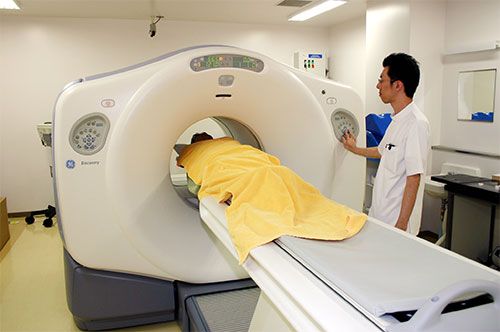
A PET mainly creates images based on metabolic function activity information and a CT creates images based on morphological information such as object location, shape and size. The greatest advantage of a PET-CT machine is that it can take superimposed images (fusion images) of a PET image and a CT image at the same time.
The superimposition of two images with the PET-CT machine drastically improves diagnostic precision by permitting the simultaneous visualization of cancerous tumors and metastatic lesions in the same position as the organs. In addition, the physical burden to the patient is reduced as both the PET and CT scans can be conducted concurrently.
In August 2005, we were the first hospital to introduce the TomoTherapy radiotherapy system in Japan, in an effort to further strengthen our diagnostic and treatment capacities.
Together with TomoTherapy, the PET-CT machine, which contributes significantly in the detection and treatment of cancer, is invaluable in determining the direction of future treatment.
Characteristics of a PET Scan
A 256-slice CT is a state-of-the-art CT machine that can take up to 256 tomographic images at a time at high speed. The CT provides three-dimensional tomographic images with higher precision and is a minimally invasive, patient-friendly diagnostic machine. We introduced the 256-slice CT in our hospital in 2016.
Conventionally, it was considered difficult to generate CT scan images of blood vessels and organs that are in constant motion such as a coronary artery. Therefore, for the diagnosis of coronary artery disease such as angina pectoris and myocardial infarction, we used to perform relatively physically burdensome cardiac catheter tests.
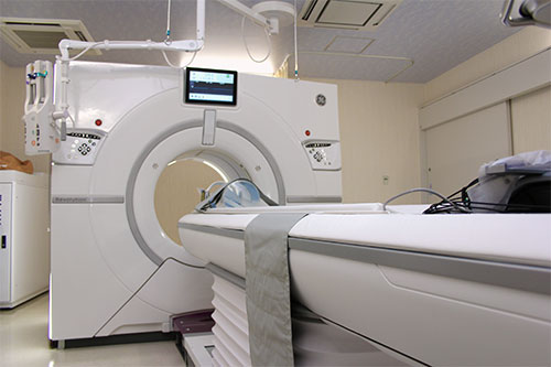
The 256-slice CT can scan a range of 16 cm per rotation (0.28 seconds). The 256-slice CT not only has the capacity to take conventional images but has also significantly improved the capacity to visualize arteries using contrast material.
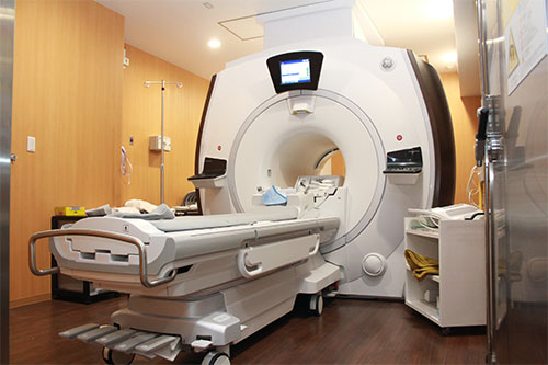
A 3-tesla MRI generates a magnetic field intensity that is twice the strength of a 1.5-tesla MRI, enabling the machine to produce higher-quality and more stable images. We were the second hospital in Japan to introduce the machine for clinical purposes.
TomoTherapy is a machine that is attracting worldwide attention as a state-of-the-art radiotherapy system for cancer treatment. We were the first hospital to introduce it in Japan.
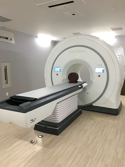
Compared to conventional radiotherapy machines, the radiation dose that TomoTherapy delivers to the areas surrounding a cancer lesion is overwhelmingly smaller and we expect TomoTherapy to minimize adverse effects to patients. TomoTherapy can treat cancer lesions of the whole body such as brain tumors, lung cancer, throat cancer and liver cancer. TomoTherapy can also deliver radiation to the whole bone marrow while avoiding brain and internal organs.
TomoTherapy has significantly expanded the range of radiotherapy, as it can not only deliver pinpoint radiation to small cancer lesions as done with gamma knives and cyber knives but also handle complicated cancer lesions and treat multiple lesions at a time.
PEM
PEMGRAPH: Positron emission tomography exclusively for the breasts (Furukawa Scintitech Corporation)
AG
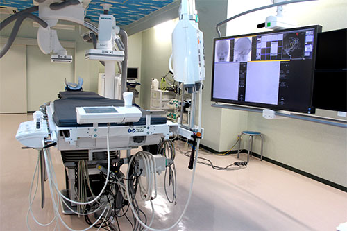
Artis Q BA Twin: Angiography (Siemens Healthineers)
Mammography
Senographe DS LaVerite: Digital mammography (GE Healthcare)
Next Generation Sequencer
MiSeq: Gene sequencer (Illumina)